 An official website of the United States government




In Situ Hybridization
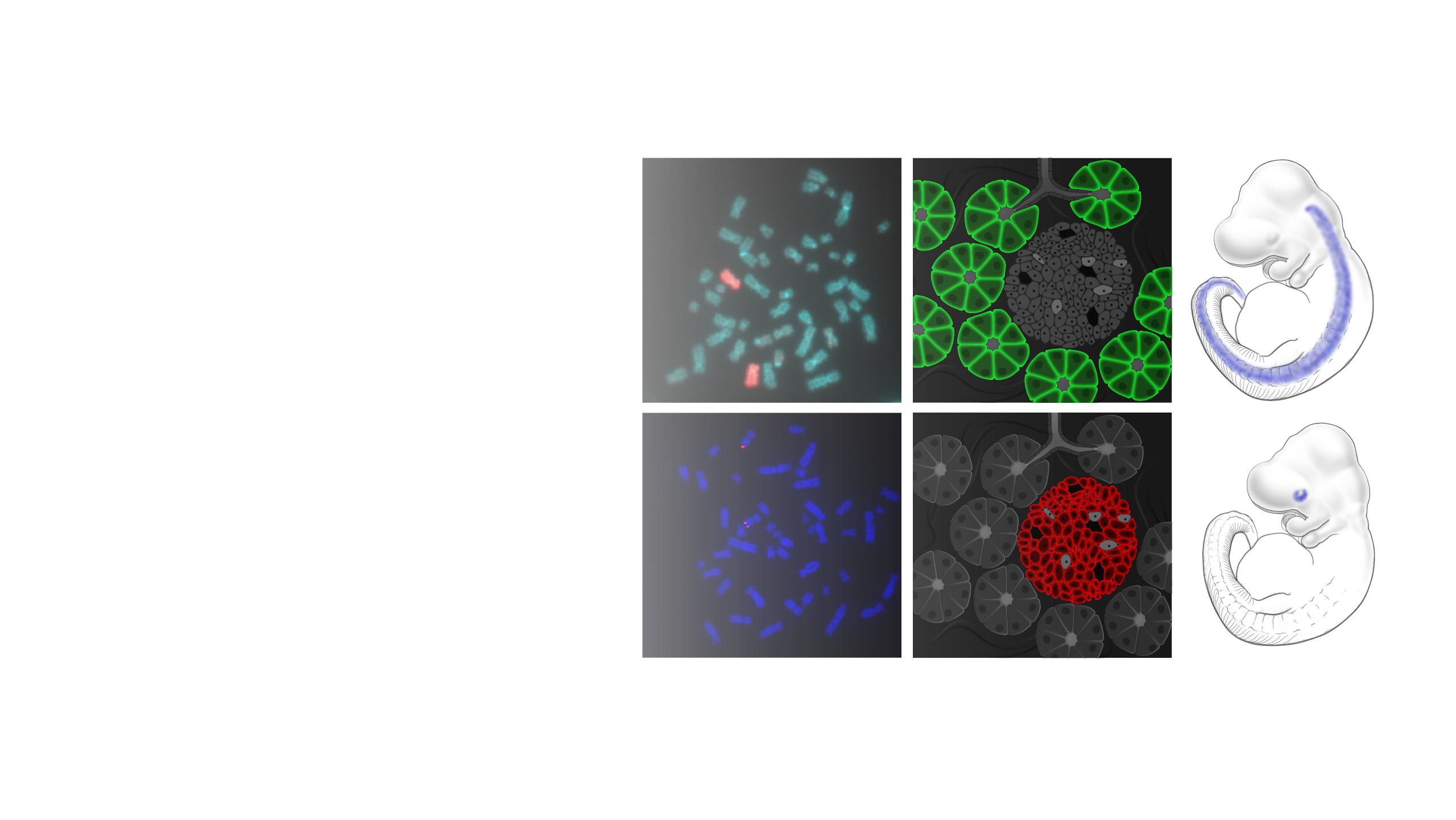
In situ hybridization is a laboratory technique used to localize a sequence of DNA or RNA in a biological sample. In this technique, a biological sample consisting of tissue sections, cells or chromosomes from an individual is affixed to a glass slide and then exposed to a “probe”—a small piece of single-stranded DNA tagged with a chemical or fluorescent dye. The labeled probe finds and then binds to its matching sequence within the biological sample. The location of the bound probe can then be seen with the use of a microscope.
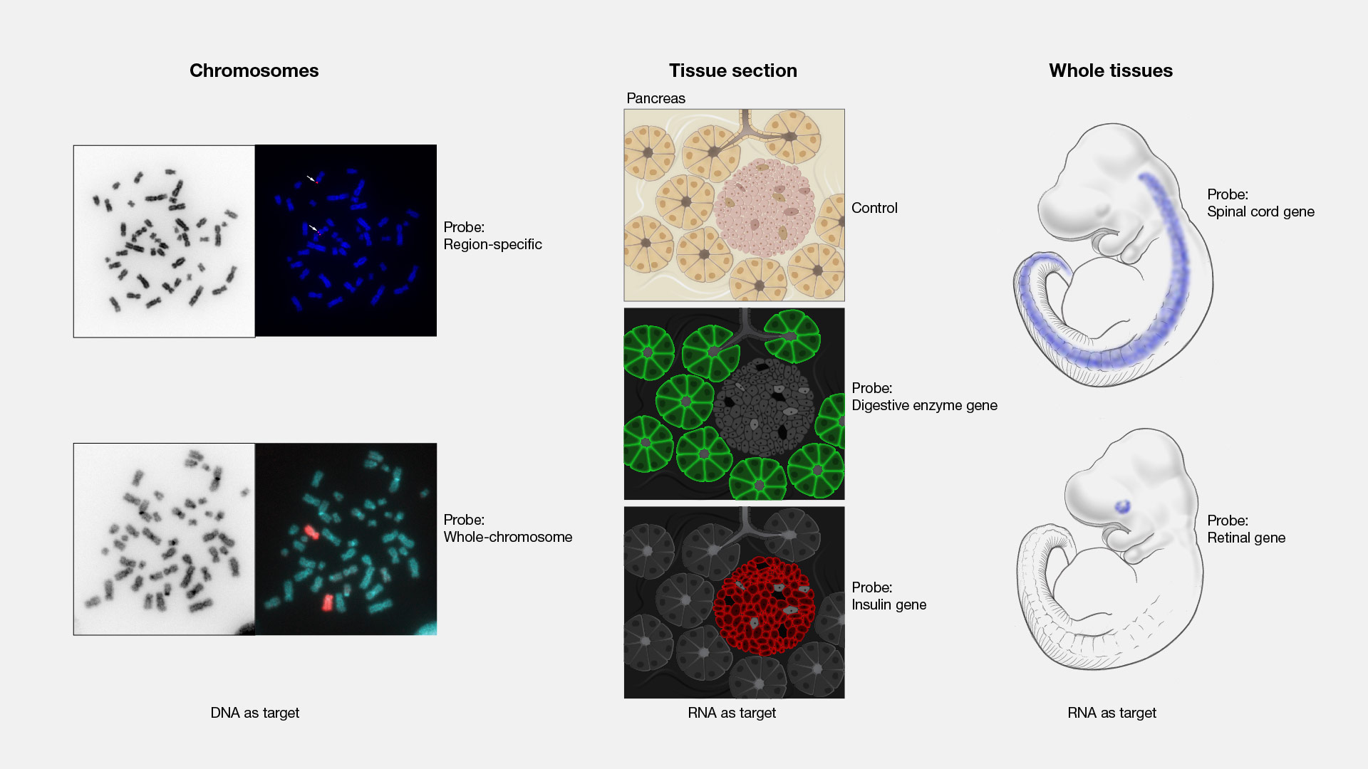
In situ hybridization. When I was a medical student, one of the hot and newly clinically available tests to look for genetic conditions in patients we were seeing was based on what we called FISH. This stands for fluorescence in situ hybridization. I remember seeing a patient at a clinic with one of the people who inspired me to become a geneticist. He was excited that we could do something called sub [inaudible] FISH testing for this patient. This means that we looked at places on the ends of each chromosome using different FISH probes.

Clinical Director
Office Of The Clinical Director
Messenger RNA (mRNA) Fluorescence In Situ Hybridization (FISH) Chromosome Hybridization Deoxyribonucleic Acid (DNA) Ribonucleic Acid (RNA) Probe